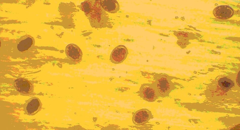
Worms under a microscope: what their eggs and parasites themselves look like

insect eggs under microscopeMany people are interested in the question of what the eggs look like, since it is not uncommon to become infected with the parasite. Infection usually occurs when eggs enter the body. This can occur due to dirty hands, food, and contact with feces and pet hair. If a parasitic infection is suspected, people will try to self-test their stool for eggs. But the eggs are not visible to the naked eye; they are so tiny that they can only be detected when stool is analyzed.

roundworm infection

Roundworm infection can occur when eating unwashed vegetables and fruits, poorly fried meat and fish. It is possible to get infected through dirty hands, especially in children. The worm's habitat is the human intestineRoundworm eggs can only be seen under a microscope. Their size is very small (about 0. 07 mm). Adult worms are also rarely seen in feces. Only after taking anthelmintics will the dead insect particles be expelled from the intestines. They look like translucent elongated inclusions.Only microscopic examination of stool can help determine the presence of roundworm eggs. The eggs are yellow and the shell is covered with nodules. Sometimes an embryo can be seen inside the fertilized egg. They are highly resistant to environmental influences and can persist outside the human body for many years.

roundworm eggs

Since it is difficult to detect traces of the presence of roundworms in the body, you should be alert to the symptoms of roundworm invasion: sudden increase in body temperature;
  • rash;
  • Choking and coughing (sometimes bloody);
  • muscle spasms;
  • joint pain.
These manifestations are related to the effects of roundworm allergens on the body. If you notice such symptoms, you will need to do a stool test to look for eggs.

Where to go if worms are suspected?

If you suspect a worm infection, you must make an appointment with an infectious disease specialist. In its early stages, helminthiasis has no specific symptoms, making it difficult to suspect that you or a loved one has worms. Usually, patients complain of mild discomfort: indigestion, headache, apathy.If symptoms do not go away within a week or if the condition returns regularly (for example, feeling unwell every 3-4 months), you should talk to your doctor. Episodes of poor health may be related to the migration of parasites.

pinworm infection

Pinworms can be contracted through casual contact with a sick person (through sharing objects, shaking hands). People are often infected by cats and dogs; the eggs live in their pets' fur. Children are particularly susceptible to this disease. Children can contract these parasites in kindergarten or from animals. Pinworm eggs can be found on any object the patient has come into contact with. They can be found under fingernails, on toys, bedding and underwear. Therefore, it is very easy to get infected with pinworms.

pinworm eggs

Pinworms can cause a disease called enterobiasis. Signs of infection include:
  • Itching in the rectal exit area;
  • diarrhea;
  • nausea;
  • Sudden weight loss;
  • Flatulence.
Pinworm eggs are not passed in the feces. Parasites multiply and lay eggs in the anal area, causing itching. To detect the presence of these worms in the body, the skin of the anus is scraped and the material obtained is examined microscopically. Such an analysis is often required when a child enters kindergarten. Gua sha is done in the morning before bathing the child to avoid washing away the parasite eggs. Perform triple analysis over several days. Pinworm eggs look like rectangular white grains under a microscope.Adult worms can be found in the feces of children and adults. These are small white bugs about 0. 5-1 cm long, with one end of the body being pointed.

Folk remedies for treating parasites

For dilofilariasis, folk remedies can be used only after consulting a doctor. They should not replace medical treatment but should only complement it. The most commonly used recipe is pumpkin seeds.Pumpkin seeds have adverse effects on many worms, including tapeworms. They contain cucurbitacins, a substance that destroys parasites. Grind the seeds with a coffee grinder or blender and dilute with water to form a paste. Adults require 300 grams of seeds and children 50 to 100 grams. The prepared product is consumed on an empty stomach for 1 hour in the morning. You should not have breakfast after this. After 3 hours, you need to take laxatives, and after another 30 minutes, you need to take an enema. When parasites come out of stool, they need to be checked. Pay attention to whether there is a head on one end of the body. If it is not present, then this means that only fragments have come out and the parasite will be able to regrow the body and release eggs. In this case, the treatment process must be repeated.

Whipworm

This parasite is quite rare in central my country. Whipworms often live in southern areas because the eggs of this worm like warmth. Most infections occur in rural areas.Whipworm eggs live in the soil. Infection occurs through hands, contaminated soil particles, and improperly washed vegetables and fruits.As a result of infection, a disease occurs - trichotillomania. Whipworms live in the intestines. The worm feeds on human blood and can cause anemia and severe abdominal pain.

whipworm eggs

The eggs of the parasite are passed in the feces, but they are so small that they are not visible even under a microscope. Only in very severe cases may eggs be detected in a stool test. They are shaped like barrels and are brown in color. The eggs have holes on both sides.What do worms in feces look like? Since whipworms cannot survive outside the body for very long, it is difficult to detect live whipworms in feces. Only with deworming treatment will you be able to find dead white worms in your stool.To diagnose trichuriasis, the rectum and sigmoid colon need to be examined using special equipment (sigmoidoscopy). In this way, the accumulation of parasites in the intestines can be detected. Because whipworm eggs are protected by a dense shell, treating the infection takes a long time.

Diagnosis of helminthiasis

When diagnosing many helminth infections, a stool examination is performed first. If you notice black spots in your stool or white worms in your stool, you should get this test done as soon as possible.However, it's not just poop with black spots that is a sign of synergistic programming. Often, even eggs that are invisible to the naked eye can be easily identified under a microscope. Stool diagnosis can be made more accurately by detecting worm DNA particles using PCR technology.If a person has a lot of black spots in their stool, other diagnostic methods include the following:
  • Scrape from the area near the anus;
  • Use ELISA, PCR, RNGA and other methods for blood testing;
  • Be sure to do blood biochemistry and CBC;
  • To determine the location of the parasite, ultrasound, MRI, and CT are performed in some cases;
  • In order to diagnose the migratory stages of the worms, X-rays are taken.
For some forms of helminthiasis, tests of sputum, rectal mucus, urine, and gallbladder contents may be done. Endoscopy is also sometimes used for diagnosis.

Trichinella spiralis

This is one of the most dangerous types of roundworms. Trichinella spiralis parasitizes human muscles. Severe infections can sometimes lead to death.Trichinella spiralis enters the body through the consumption of poorly processed meat from wild and domestic animals. Worms are only destroyed at very high temperatures (about 80°C). Worms may be present in bacon or bacon; this treatment will not kill the larvae.Undercooked meat may cause infectionParasite eggs cannot be detected in humans. The female Trichinella spiralis carries the eggs into her body, and the larvae are born. These are ovoviviparous worms. It is impossible to detect Trichinella spiralis in feces. Newly born larvae immediately bypass the intestines and enter the blood and lymph fluids. The larvae soon die in the feces.Typically, the disease is diagnosed when the parasite successfully enters the muscle. In this case, a person suffers from the following symptoms: muscle pain;
  • swelling;
  • Fever status (high temperature, pain, discomfort);
  • Irregular bowel movements with constipation or diarrhea.
To detect invasion, blood tests and serology tests are required. This is the only way to detect Trichinella spiralis in your body.An article for patients suffering from a medically diagnosed disease. It is not intended to replace a doctor's appointment or be used for self-diagnosis.

tapeworm

The human body contains only immature tapeworm eggs. They are excreted through feces and enter the external environment. If the wastewater is left untreated, the eggs end up in bodies of water where they begin to develop. First, they end up inside freshwater crustaceans. Fish in reservoirs become infected with tapeworms when they eat small crustaceans. The worms become infected when a person eats poorly fried, infected fish or raw pike caviar from a freshwater body.

tapeworm eggs

The occurrence of dilofilariasis is manifested by the following symptoms: abdominal pain;
  • nausea and vomiting;
  • Intestinal problems (constipation or diarrhea);
  • Loss of appetite or excessive hunger.
What do tapeworm worms look like? This is a large parasite that can reach 10 m in length. In the feces, only individual living parts (segments) of the worms can be found; they look like long (30 cm to 3 m) white ribbons. It should be removed from the feces using forceps, transferred to a clean container and given to a parasitologist or infectious disease specialist for analysis.Microscopic examination of stool can detect tapeworm eggs. Their size is approximately 0. 07 mm. The eggs look like yellowish oval structures covered with a thick shell. The egg has a cap on one end and a bump on the other end.Worm larvae can be passed in the feces but are not dangerous. Infected people or animals cannot become infected with diye filariasis. Infection occurs only through consumption of fish.

Harm to the body

Dilofilariasis occurs when large tapeworms enter the intestines. Worms primarily affect the gastrointestinal tract. Inflammation and ulcers can form on the intestinal wall where the worms attach. If there are multiple parasites in the body instead of just one, they can clog the intestinal lumen, causing an obstruction. Worms constantly irritate the walls of the gastrointestinal tract, causing disturbances in the digestive process. In addition, it poisons the human body through waste products, thereby causing allergies. Severe anemia and vitamin B12 deficiency can occur when the parasite remains in the body for an extended period of time.

Cattle and pork tapeworms

People become infected with these types of parasites by eating poorly processed livestock meat. Worm segments are passed in the patient's stool. In the external environment, the segments pass through the soil and lay eggs, which contain larvae. The eggs are then ingested by pets. When a person eats contaminated beef or pork, they become infected with the bovine or pork tapeworm. To kill tapeworms, boil or fry meat for at least 30 minutes.

bull tapeworm

The bovine tapeworm causes taeniasis, and the pork tapeworm causes taeniasis. The symptoms of these diseases are similar: abdominal pain;
  • persistent hunger;
  • nausea and vomiting;
  • weakness;
  • lose weight;
  • diarrhea;
  • Itching in the anal area as the segments fall off.
The worms in the patient's stool are segmented. They look like light streaks about 1-2 cm long. The segment of the pork tapeworm is longer and consists of 3 segments.When stool is analyzed, tapeworm eggs (taenia ova) can be detected. They are round in shape and have a dense shell containing an embryo.Pork tapeworm can be contracted through dirty hands and there is no intermediate host. The fragments excreted in the patient's stool are dangerous. They can enter the body from contaminated soil. In this case, the solium tapeworm larvae multiply in the human body and cause a serious disease called cysticercosis. This is a very dangerous invasion. The larvae enter the brain, spinal cord, eyes, heart and lungs, causing severe damage. In cysticercosis, the segments and eggs are not passed in the feces. The disease can only be detected through serological blood tests and cerebrospinal fluid analysis.

Classification

Modern medicine divides worms that parasitize the human body into the following categories: Luminal. These worms live in the intestinal lumen. These include broad tapeworms, dwarf tapeworms, bovine tapeworms, hookworms, pinworms, whipworms, roundworms, etc.Fabric. These worms choose muscle and lung tissue as their habitat, as well as organs such as the pancreas, liver, and brain.Depending on the specific location of the organization's worm, the intrusion may have the following names:
  • Filariasis. Parasites live in lymph nodes
  • Cysticercosis. Areas of the brain affected by worms
  • Hydatid disease. Worm infection diagnosed in liver
  • Paragonimiasis. Parasites live in the lungs

Fluke

Among trematode worms, cat flukes (liver flukes) are most commonly found in humans. The habitat of the eggs is fresh water. From there, the parasite makes its way into the shellfish and then into the fish. Cats and people become infected with flukes by eating poorly prepared freshwater fish and contaminated water. Sick cats pose no danger to humans.

Burbot liver has parasites

Most commonly, fish of the family Cyprinidae are infected. Salting or smoking will not kill the parasites. The product requires considerable heat treatment. Flukes can be contracted by accidentally swallowing water from a pond or river. There are known cases of intrusion occurring after beds have been watered with contaminated water.Cat flukes attack the liver. Right abdominal pain, nausea, vomiting, and fever. During the physical examination, organ enlargement was found.Adult worms are not passed in the feces. What do fluke eggs look like under a microscope? When the stool is examined, a clear oval shape with a golden outer shell can be seen. The eggs have a plug on one side that opens when the larvae hatch. For diagnostic purposes, an antibody blood test or enzyme-linked immunosorbent assay is also performed.

How to tell if there are bugs?

It is not possible to independently determine whether a worm infection is present. In its initial stages, the disease may actually be asymptomatic. The patient feels no pain; the immune system suppresses the disease-causing effects of toxins and allergens for a period of time. Typically, the condition begins to worsen during larval migration or as the worm population increases. The more severe the infection (i. e. the more parasites there are), the more symptoms will occur.However, the asymptomatic invasion process is dangerous - the patient infects others, and his health gradually deteriorates. In order to detect this disease, it is necessary to go to the hospital regularly for preventive examinations. As part of prevention, therapists perform worm testing at least once a year. If you live in an endemic area - every six months.

What can the naked eye see?

Due to the very small size of some parasites, it is not possible in all cases to detect their presence in the body simply by the presence of eggs in the stool. Some parasites are very tiny in size and hide within the body without revealing their presence. Furthermore, they are not always localized to the gut but are able to migrate throughout the body. Therefore, to diagnose parasitic infections, they use serological tests based on antigen-antibody immune responses.All parasites look different, have their own specific development cycles, different symptoms of infection, and differences in treatment options. However, there are many symptoms that may indicate a parasitic infection in the body:
  • rapid weight loss;
  • Intestinal disorders: Diarrhea replaces constipation;
  • Severe itching of the anus;
  • Rash of unknown cause;
  • stomach ache;
  • flatulence;
  • Loss of appetite;
  • An inexplicable craving for sweets;
  • Sometimes adults cannot control their appetite;
  • Frequent colds due to reduced body resistance.